Air Embolism: Causes, Recognition, and Emergency Management
Venous and arterial air embolisms are rare but life-threatening complications associated with central venous catheter placement, surgery, or diving accidents. This article covers pathophysiology, clinical signs, the Durant maneuver, and the role of hyperbaric oxygen therapy.

Author: Dr. med. univ. Daniel Pehböck, DESA
Specialist in Anesthesiology and Intensive Care Medicine, AHA-certified ACLS/PALS Instructor, Course Director Simulation Tirol
Reading time approx. 9 min
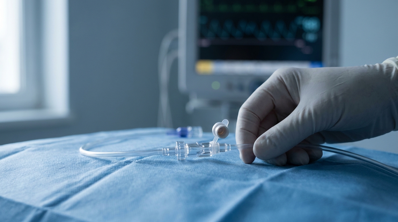
Air embolisms are among those emergencies that rarely occur in clinical practice but can rapidly become fatal if diagnosis and treatment are delayed. The spectrum ranges from clinically inapparent microembolisms to fulminant cardiovascular collapse. The pathophysiology differs fundamentally between venous and arterial air embolism – and so does the therapeutic approach. For emergency physicians, anesthesiologists, and intensive care personnel, a solid understanding of the mechanisms, clinical recognition, and immediate countermeasures is essential. This article systematically summarizes the relevant aspects and provides clear management recommendations.
Definitions and Epidemiology
An air embolism occurs when air – or another gas – enters the venous or arterial vascular system and causes a hemodynamically significant obstruction. The distinction between venous air embolism (VAE) and arterial air embolism (AAE) is clinically crucial, as causes, pathophysiology, and treatment differ considerably.
Venous Air Embolism (VAE)
VAE is the more common form. Air enters the right heart and pulmonary vasculature through open veins. The incidence depends heavily on the clinical context:
- Neurosurgical procedures in the sitting position: Incidence up to 80% when sensitive detection methods are used (precordial Doppler ultrasonography)
- Central venous catheter (CVC) insertion and removal: Estimated incidence of subclinical embolisms at 1–2% of all catheterizations
- Laparoscopic procedures: Rare, but documented during insufflation into open venous plexuses
- Obstetrics: Particularly during cesarean section and uterine atony
Arterial Air Embolism (AAE)
AAE is less common but generally more severe. Mechanisms include:
- Paradoxical embolism via a patent foramen ovale (PFO) – prevalence in the general population approximately 25–30%
- Direct arterial insufflation during arterial catheterization, cardiopulmonary bypass, or penetrating injuries
- Pulmonary barotrauma in diving accidents (arterial gas embolism, AGE)
- Crossover during massive VAE, when the filtering capacity of the pulmonary vasculature is exceeded
Pathophysiology
Venous Air Embolism
The pathophysiological consequences of a VAE depend on the volume and rate of air insufflation. The lethal dose in adults is cited in the literature as 3–5 ml/kg, though the infusion rate is critical: slow delivery of small amounts is absorbed by the pulmonary microcirculation, while rapid bolus administration can be lethal even at lower volumes.
The mechanism of hemodynamic decompensation proceeds as follows:
- Air accumulation in the right ventricle: The air forms a compressible foam that drastically reduces the ejection fraction of the right ventricle.
- "Air-lock" phenomenon: A large air bolus blocks the right ventricular outflow tract and prevents any forward flow into the pulmonary artery.
- Pulmonary vasoconstriction: Air in the pulmonary arteries triggers reflex vasoconstriction, further increasing pulmonary vascular resistance.
- Right heart failure: The acute pressure overload on the right ventricle leads to dilation, leftward septal deviation, and consequent decrease in left ventricular output.
- Cardiovascular collapse: Without intervention, obstructive shock ensues.
Additionally, activation of the coagulation cascade and complement cascade occurs at the air-blood interface, promoting a secondary inflammatory response with endothelial damage, capillary leak, and pulmonary edema.
Arterial Air Embolism
In AAE, air bubbles enter the systemic circulation and obstruct end-arterial vascular territories. The consequences depend on the affected vascular territory:
- Cerebral AAE: Focal neurological deficits, loss of consciousness, seizures – clinically often indistinguishable from an ischemic stroke
- Coronary AAE: ST-segment changes, arrhythmias, cardiogenic shock
- Mesenteric AAE: Abdominal pain, signs of ischemia
- Cutaneous AAE: Livedo reticularis as a pathognomonic sign
The air in the arterioles causes not only mechanical obstruction but also endothelial damage through oxidative stress and a local inflammatory response, which extends the degree of ischemia beyond the pure obstruction time.
Risk Factors and Clinical Scenarios
CVC Insertion and Removal
CVC-associated air embolism is a classic and preventable event. Air typically enters during:
- Disconnection of the infusion system
- Uncapped catheter lumens during manipulation
- CVC removal without adequate positioning and closure of the puncture site
- Spontaneous breathing with negative intrathoracic pressure in the upright position
Prevention is particularly effective here: Trendelenburg positioning, Valsalva maneuver, airtight connections, and consistent capping of all lumens are simple measures with high efficacy.
Surgical Procedures
Procedures are particularly high-risk when non-collapsible veins (e.g., dural sinuses, diploic veins of the calvarium) are opened in the surgical field and are located above heart level. The sitting position in posterior fossa surgery is the classic high-risk scenario.
Diving Accidents
During ascent from depth, gas in the lungs expands according to Boyle's law. If the air is not exhaled – for example due to panic, laryngospasm, or obstructive pulmonary disease – alveolar rupture can occur. The air enters the left atrium directly via the pulmonary veins and from there into the systemic circulation. An AGE can occur during an ascent from as little as 1–2 meters depth if air is held.
Clinical Signs and Diagnostics
The clinical presentation is variable and depends on the volume of embolized air and the affected vascular territory.
Signs of Venous Air Embolism
- Early signs: Sudden drop in end-tidal CO₂ (etCO₂), tachycardia, hypotension
- Auscultation: Characteristic "mill-wheel murmur" – a continuous, churning sound over the precordium; however, this is a late sign indicating an already massive embolism
- Hemodynamics: Rise in CVP, drop in cardiac output, pulseless electrical activity (PEA) or asystole in massive events
- Pulse oximetry: SpO₂ drop due to ventilatory dead space
- Capnography: Abrupt etCO₂ drop – one of the most sensitive and earliest signs on monitoring
Signs of Arterial Air Embolism
- Neurological: Acute hemiparesis, aphasia, seizures, loss of consciousness
- Cardiac: ST elevations/depressions, malignant arrhythmias
- Cutaneous: Livedo reticularis, mottled skin
- Retinal: Air bubbles visible on fundoscopy (pathognomonic, but rarely examined in an emergency)
Diagnostic Modalities
- Transesophageal echocardiography (TEE): Gold standard for intraoperative detection – can identify individual air bubbles in the right heart
- Precordial Doppler: Highly sensitive, capable of detecting as little as 0.01–0.05 ml/kg of air
- Transthoracic echocardiography (TTE): Rapidly available in emergencies, shows air in the right ventricle and paradoxical septal motion
- CT thorax: Can demonstrate intracardiac and intravascular air, but not a primary diagnostic method in the acute setting
Emergency Management
Treatment must begin immediately and is guided by the severity and type of embolism.
Immediate Measures for Venous Air Embolism
- Stop the air entry: Identify and seal the entry point – this is the most important and most urgent measure.
- Left lateral decubitus with head-down tilt (Durant maneuver): The patient is placed in the left lateral position, ideally with 15–30° head-down tilt (Trendelenburg). The goal: the air in the right ventricle rises to the lateral portion of the right ventricle and clears the right ventricular outflow tract (RVOT). This improves forward flow into the pulmonary artery and prevents complete air-lock.
- 100% oxygen: Maximize FiO₂ to 1.0. Oxygen accelerates nitrogen resorption from the air bubbles by increasing the diffusion gradient (analogous to the principle of denitrogenation). Simultaneously, hypoxemia is treated.
- Volume resuscitation: Aggressive fluid therapy to increase venous return and compress the air bubbles.
- Aspiration via CVC: If a central venous catheter with a correctly positioned tip (at the junction of the superior vena cava/right atrium) is in place, an attempt can be made to aspirate air. The success rate is limited, but in individual cases it can be lifesaving.
- Vasopressors: For persistent hypotension, norepinephrine or epinephrine is indicated to maintain systemic perfusion pressure.
- CPR in cardiac arrest: For PEA or asystole, follow the standard AHA algorithm, but maintain the left lateral decubitus position if possible. Chest compressions can help fragment the air bolus and transport the smaller bubbles into the pulmonary vasculature, where they are more readily absorbed.
The Durant Maneuver in Detail
The maneuver, named after the American surgeon Thomas Durant, is one of the few specific positioning interventions in emergency medicine. The physiological rationale:
- In the supine position, the RVOT lies anteriorly and thus uppermost – air accumulates there and blocks the outflow tract.
- In the left lateral decubitus position, the heart rotates, the RVOT shifts laterally and inferiorly, and the air rises into the now higher-lying lateral portion of the right ventricle – away from the outflow tract.
- The head-down tilt additionally prevents further air entry through open veins above heart level.
The evidence for the Durant maneuver is based primarily on animal experimental data and case reports. Randomized studies are lacking – which is not surprising given the emergency nature and rarity of the event. The maneuver is nevertheless considered an established standard, as it is low-risk and pathophysiologically plausible.
Management of Arterial Air Embolism
AAE requires additional measures:
- Flat positioning or slight head-down tilt: A previously recommended steep Trendelenburg position is no longer generally recommended, as it can worsen cerebral edema.
- 100% oxygen: As with VAE, but here with particular emphasis on accelerating bubble resorption in end-arterial territories.
- Hyperbaric oxygen therapy (HBO): For AAE – especially the cerebral form and AGE after diving accidents – HBO is the definitive therapy.
- Seizure control: For cerebral AAE with seizures, benzodiazepines (e.g., midazolam 1–2 mg IV titrated) are the first-line treatment.
- Avoidance of nitrous oxide: In anesthesia, nitrous oxide must be discontinued immediately, as it diffuses into air bubbles and increases their volume (35-fold solubility compared to nitrogen).
Hyperbaric Oxygen Therapy (HBO)
HBO is the only causative therapy that directly reduces the volume of air bubbles. The mechanisms of action are:
- Boyle's law: At a pressure of 2.8 ATA (typical treatment protocol), the bubble volume is compressed to approximately one-third.
- Accelerated nitrogen elimination: Under hyperbaric oxygen, the diffusion gradient for nitrogen from the bubble into the surrounding tissue increases massively, leading to rapid dissolution.
- Tissue oxygenation: Despite microvascular obstruction, the high pO₂ enables adequate oxygen diffusion into the ischemic tissue.
- Anti-inflammatory effect: HBO reduces leukocyte adhesion to the damaged endothelium and diminishes reperfusion edema.
Indications for HBO in Air Embolism
- Cerebral arterial air embolism – regardless of the cause
- Arterial gas embolism (AGE) after diving accident
- Venous air embolism with persistent neurological deficits (suggestive of paradoxical embolism)
- Any air embolism with treatment-refractory hemodynamic instability
Treatment should be initiated as early as possible, ideally within 4–6 hours. Even with later presentation – up to 24 hours and beyond – a therapeutic attempt may be justified, particularly in the presence of neurological deficits.
In Austria, hyperbaric chambers are available at multiple locations. Transport must be carried out with continued 100% oxygen administration, ideally by ground – or, in the case of helicopter transport, with cabin altitude at ground level to avoid further bubble expansion due to pressure reduction.
Prevention
The best treatment for air embolism is prevention. Key preventive measures include:
- CVC management: Trendelenburg positioning during insertion and removal, capping of all lumens, use of Luer-Lock connections, Valsalva maneuver or having the patient bear down during disconnection
- Intraoperatively: Flooding the surgical field with saline when venous sinuses are open, vigilance during operations above heart level, monitoring with precordial Doppler or TEE during high-risk procedures
- Infusion systems: De-airing all infusion lines, use of air detectors in automated infusion systems
- Diving medicine: Never hold your breath during ascent, slow controlled ascent, adherence to decompression stops
Differential Diagnoses
In cases of sudden intraoperative or catheter-associated circulatory collapse, the following differential diagnoses must be considered:
- Pulmonary embolism (thrombotic)
- Anaphylaxis (drug-related, latex)
- Tension pneumothorax
- Pericardial tamponade
- Myocardial infarction
- Local anesthetic systemic toxicity (LAST)
Capnography can help differentiate air embolism from anaphylaxis: in air embolism, etCO₂ drops abruptly, while in anaphylaxis it often remains preserved or is altered by bronchospasm.
Summary: Algorithm for Suspected Air Embolism
- Seal the entry point – clamp the CVC, flood the surgical field, remove the catheter
- 100% oxygen – FiO₂ 1.0, discontinue nitrous oxide immediately
- Durant maneuver – left lateral decubitus, 15–30° head-down tilt
- Volume resuscitation – aggressive, crystalloid and/or colloid
- Aspiration via CVC – if in place, attempt air aspiration
- Vasopressors – norepinephrine/epinephrine as needed
- CPR – in cardiac arrest per AHA algorithm
- Evaluate HBO – especially for neurological deficits or AAE
- Monitoring – TEE/TTE, capnography, close hemodynamic monitoring
Practical Training
Air embolism is a prime example of an emergency where theoretical knowledge alone is not enough. The Durant maneuver, rapid identification of the entry point, and coordinated teamwork under time pressure must be practiced before they are needed in a real situation. In the Emergency Physician Refresher Course by Simulation Tirol, you train exactly these rare but critical scenarios on realistic simulation models – with structured debriefing and the opportunity to internalize algorithms under controlled stress. Because especially in emergencies that you may encounter only once in your career, preparation determines the outcome.
More Articles
Recognizing Agonal Breathing: How to Correctly Interpret Gasping
Agonal breathing is frequently misinterpreted as normal breathing, delaying the start of resuscitation. This article explains its characteristics, how to differentiate it from physiological breathing, and the consequences for first responders and healthcare professionals.
Acute Dyspnea: Differential Diagnosis and Immediate Management
Dyspnea has numerous causes ranging from asthma to heart failure to pulmonary embolism. This article presents a systematic approach to differentiation based on history, clinical examination, auscultation, and point-of-care diagnostics.
Acute Hypoglycemia: Emergency Management in Adults and Children
Threshold values, symptom recognition, oral glucose vs. IV dextrose vs. glucagon – with separate dosing for adult and pediatric patients. A common emergency presentation systematically reviewed.